The Dental X-Ray Safety: Radiation Levels and Frequency Explained
Dental X-rays are an essential diagnostic tool in modern dentistry, yet many patients express understandable concerns about radiation exposure. At our practice, patient safety remains our top priority. This comprehensive guide explains dental X-ray safety, radiation levels, recommended frequency, and why these low-dose images play a vital role in maintaining your oral health.
Schedule Your Safe Digital X-Ray Today – Call Now!
Whether you're worried about cumulative radiation or simply want to understand the process better, this article provides clear, evidence-based information to help you feel confident about necessary dental imaging.
Understanding Dental X-Rays and Patient Concerns
Many people associate X-rays with high radiation from medical procedures or airport scanners. However, dental X-rays use significantly lower doses than most other types of imaging.
Modern digital dental radiography has revolutionized safety standards. Today's equipment delivers precise images with a fraction of the radiation used in older film-based systems. This advancement allows dentists to detect hidden issues early while minimizing exposure.
Common concerns we hear include:
- Will repeated X-rays increase cancer risk?
- Are dental X-rays safe during pregnancy?
- How much radiation will I actually receive?
These are valid questions. Let's address them directly with factual data.
How Much Radiation Do Dental X-Rays Actually Use?
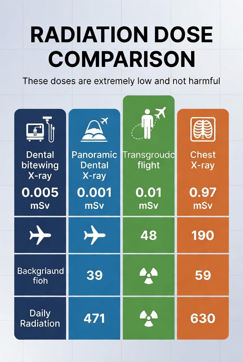
The numbers speak for themselves. Here are typical effective radiation doses for common dental X-rays:
- Bitewing X-rays: Approximately 0.005 mSv
- Panoramic X-ray: Approximately 0.01 mSv
- Full-mouth series: Usually between 0.02 – 0.05 mSv depending on technique
To put this in perspective, the average person receives about 3 mSv of background radiation per year from natural sources like cosmic rays and radon gas. A single bitewing X-ray equals roughly one to two days of normal background radiation. A panoramic X-ray is comparable to a short domestic flight.
Digital sensors have reduced radiation exposure by up to 80-90% compared to traditional film X-rays. This means safer diagnostics for every patient.
Our practice uses state-of-the-art digital X-ray systems that not only lower radiation but also provide instant, high-resolution images for accurate diagnosis.
Why Dental X-Rays Are Medically Necessary
Dental professionals cannot see everything during a visual examination. X-rays reveal critical information hidden beneath the surface:
- Cavities between teeth that visual inspection might miss
- Early bone loss from gum disease
- Infections at tooth roots or in the jawbone
- Impacted teeth or developmental abnormalities
- Abscesses and cysts
- Problems with existing dental work
Without X-rays, many serious issues would go undetected until they cause pain or require more extensive treatment. Early detection through safe imaging often means simpler, less invasive, and more affordable solutions.
Benefits of Regular Dental X-Rays
- Prevent small problems from becoming major dental emergencies
- Monitor healing after procedures
- Track progression of periodontal disease
- Support orthodontic treatment planning
- Ensure implants and crowns are functioning properly
The diagnostic value far outweighs the minimal radiation risk for most patients.
Recommended Frequency of Dental X-Rays
The frequency of X-rays depends on your individual oral health needs, age, and risk factors. General guidelines include:
- Bitewing X-rays: Typically recommended once per year for most adults with moderate risk
- Full-mouth series: Usually every 3 to 5 years
- Panoramic X-rays: Every 3-5 years or as needed for specific concerns
- Children and adolescents: May need more frequent imaging during growth phases to monitor tooth development
High-risk patients (those with history of extensive decay, gum disease, or smoking) may require X-rays more often. Low-risk patients with excellent oral hygiene might safely extend intervals.
Your dentist will always customize recommendations based on your unique situation rather than applying a one-size-fits-all approach.
Safety Measures That Protect Patients
Modern dental offices implement multiple layers of protection:
- Lead aprons and thyroid collars shield sensitive areas
- High-speed digital sensors require less radiation
- Collimators focus the X-ray beam precisely
- Strict protocols ensure proper technique and minimal retakes
- Pregnancy protocols with additional precautions when necessary
For pregnant patients, we carefully weigh benefits against risks and often postpone non-urgent X-rays, especially during the first trimester. However, when diagnostic needs are urgent, the low doses used in dentistry are considered safe with proper shielding.
Different Types of Dental X-Rays and Their Uses
Understanding the various types helps patients appreciate their targeted purpose:
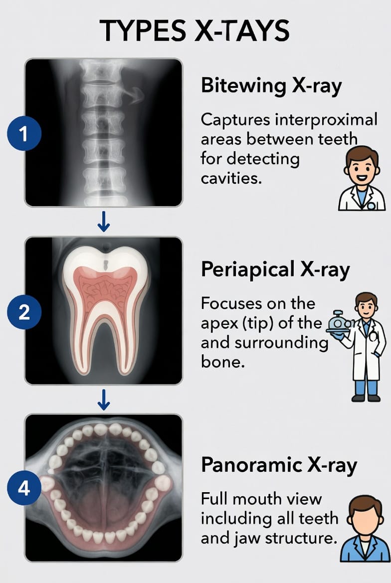
Bitewing X-Rays
These show the upper and lower back teeth in one view. They excel at detecting decay between teeth and changes in bone height.
Periapical X-Rays
These focus on one or two teeth from crown to root tip. They help diagnose root infections, abscesses, and bone changes around specific teeth.
Panoramic X-Rays
A single image captures the entire mouth, including wisdom teeth, jaw joints, and sinuses. Useful for overall assessment and surgical planning.
Cone Beam CT (CBCT)
Used in complex cases like implant planning, this 3D imaging provides detailed views while still using relatively low radiation for its diagnostic power.
Addressing Radiation Risk Myths
Scientific studies consistently show that the radiation levels from dental X-rays pose very low risk. The American Dental Association and FDA endorse current safety protocols as appropriate for diagnostic needs.
The risk from undiagnosed dental problems — such as progressing infections or undetected oral cancers — often presents a greater health concern than the minimal radiation from necessary X-rays.
Key reassurance: The technology, protocols, and professional judgment used today make dental X-rays among the safest medical imaging procedures available.
Who Should Be Extra Cautious?
While dental X-rays are safe for most people, certain groups receive additional consideration:
- Pregnant women
- Young children
- Patients with certain medical conditions
- Individuals who have recently undergone multiple medical X-rays
In these cases, your dental team will discuss alternatives when possible and take every precaution necessary.
The Future of Dental Imaging
The field continues advancing toward even lower radiation and higher precision. Artificial intelligence now helps enhance images, potentially reducing the need for multiple exposures. Digital records also allow better tracking of your imaging history across visits.
Making Informed Decisions About Your Dental Care
Understanding dental X-ray safety empowers you to participate actively in your oral health decisions. When your dentist recommends X-rays, they do so based on clinical need — not routine protocol alone.
Safe, low-dose imaging remains essential for comprehensive dental health. Modern dentistry balances diagnostic benefits with patient safety through advanced technology and personalized care.
If you have questions about dental X-rays or want to learn more about our safety protocols, we’re here to help.
Ready for your next check-up? Ask about our digital X-rays and experience the difference modern, low-radiation technology makes.
Your smile deserves the best care — safe, effective, and backed by science.
Ready for Safe & Accurate Dental Imaging? Call Now