The Dental X-Ray Frequency: How Often Is Safe and Necessary
Understanding how often you need dental X-rays is one of the most common questions patients ask during routine visits. Many wonder if frequent imaging poses risks or if skipping them could miss important issues. The good news is that dental X-rays are an essential, safe diagnostic tool when used appropriately. This comprehensive guide explains recommended frequencies, the factors that influence your personal schedule, and why these imaging protocols support long-term oral health.
Discover How Often You Need Dental X-Rays – Call Today
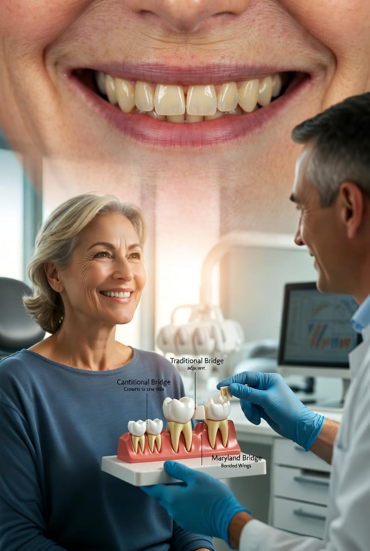
At our practice, we prioritize patient safety while delivering thorough care. Dental X-rays allow dentists to detect problems invisible to the naked eye, enabling early intervention that preserves your natural smile.
Why Dental X-Rays Matter for Your Oral Health
Dental X-rays, also known as radiographs, provide critical insights into teeth, roots, bone structure, and surrounding tissues. They reveal cavities between teeth, bone loss, impacted teeth, infections, and other conditions that routine visual exams might overlook.
Early detection through appropriate X-ray frequency prevents minor issues from becoming major dental emergencies. For instance, catching a small cavity early can mean a simple filling instead of a root canal or crown. This proactive approach saves time, money, and discomfort while maintaining your confident smile.
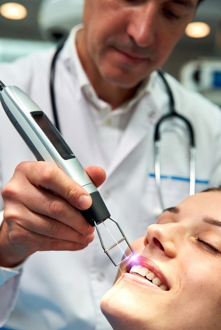
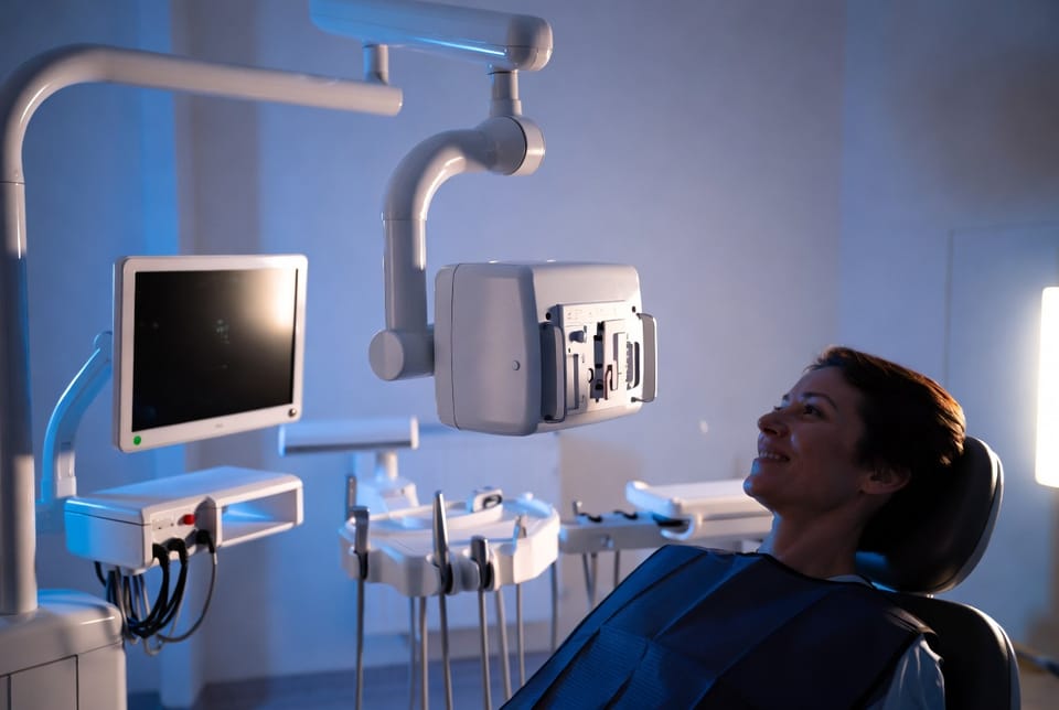
Modern digital X-ray technology has significantly reduced radiation exposure compared to older film methods—often by up to 90%. Combined with lead aprons and thyroid collars, the process prioritizes your protection without compromising diagnostic quality.
Standard Dental X-Ray Schedules for Most Patients
Dental professionals follow evidence-based guidelines tailored to individual needs. Here are the typical recommendations:
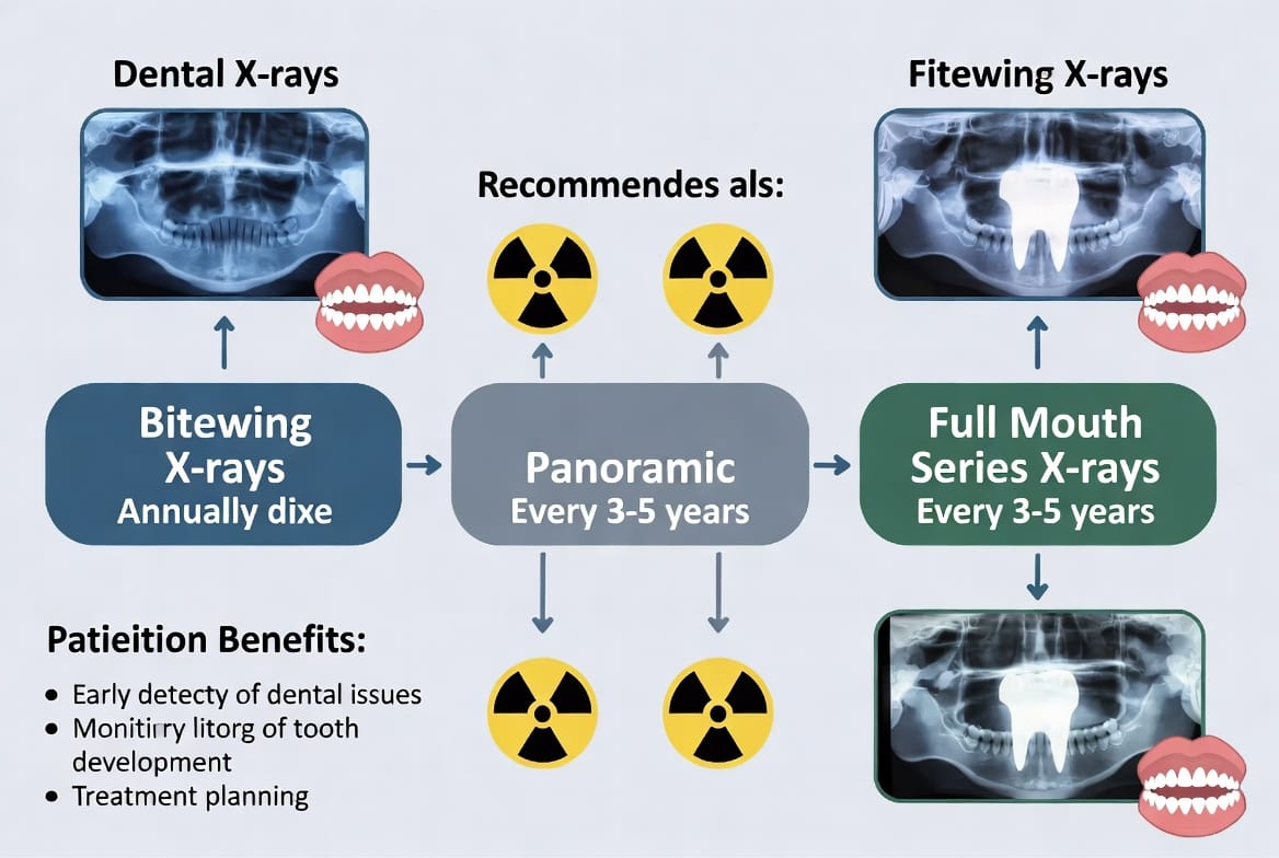
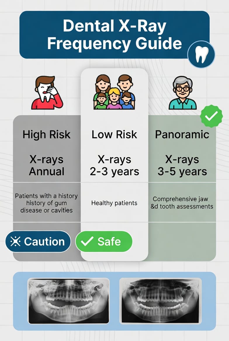
- Bitewing X-rays: Usually recommended annually for most adults. These images focus on the crowns of upper and lower teeth, making them excellent for detecting interproximal cavities (decay between teeth) and monitoring bone levels.
- Panoramic X-rays: Typically taken every 3-5 years. This single image captures the entire mouth, including wisdom teeth, jaw joints, and sinuses. It's particularly valuable for orthodontic planning, implant evaluation, and assessing overall dental development.
- Full Mouth Series (FMX): Often performed every 3-5 years for comprehensive baseline records. This set of 14-20 images provides detailed views of all teeth and supporting structures.
These intervals represent a balanced approach for patients with average risk profiles. Your dentist will adjust based on clinical findings and health history to ensure necessary imaging without excess.
How Individual Risk Factors Determine Your X-Ray Frequency
One size does not fit all when it comes to dental X-ray frequency. Dentists assess several key risk factors to create a personalized imaging plan:
- Cavity History: Patients with frequent decay may need bitewings more often—potentially every 6 months—to monitor progression and effectiveness of preventive measures.
- Age Considerations: Children and adolescents often require more frequent imaging due to rapid dental development, erupting permanent teeth, and orthodontic needs. Adults with stable oral health may extend intervals safely.
- Overall Health and Habits: Conditions like gum disease, diabetes, dry mouth (xerostomia), or smoking increase risk and may warrant more regular X-rays. Pregnancy requires special precautions, though necessary dental imaging can proceed with proper shielding.
- Previous Dental Work: Crowns, bridges, implants, or root canals benefit from periodic radiographic monitoring to ensure longevity and detect complications early.
Low-risk patients (excellent hygiene, no decay history, healthy gums) might safely reduce frequency to bitewings every 18-24 months and comprehensive imaging every 5 years.
High-risk patients (active decay, periodontal concerns, medical conditions) follow more vigilant schedules with annual or semi-annual imaging as needed.
This risk-based approach ensures you receive exactly the diagnostic support your smile requires—neither too little nor too much.
Addressing Safety Concerns: Radiation and Modern Protections
Many patients express concern about cumulative radiation from dental X-rays. Rest assured, the radiation dose from a standard dental X-ray series is extremely low—equivalent to the background radiation you receive in a few days of normal living.
Digital sensors and collimators further minimize exposure by focusing only on the necessary area. Every patient receives a lead apron and, when appropriate, a thyroid collar. These simple yet effective barriers block scatter radiation, providing peace of mind.
Organizations like the American Dental Association (ADA) and FDA endorse these protocols, confirming that diagnostic benefits far outweigh minimal risks when following recommended guidelines.
Key safety practices include:
- Using the ALARA principle (As Low As Reasonably Achievable)
- Reviewing your full health and imaging history before each series
- Employing state-of-the-art digital equipment
- Customizing frequency based on clinical necessity rather than routine alone
Benefits of Following Recommended X-Ray Schedules
Consistent, appropriate dental imaging delivers numerous advantages:
- Early Problem Detection: Identify decay, infections, or bone loss before symptoms appear.
- Treatment Planning: Provide accurate information for procedures like implants, orthodontics, or extractions.
- Progress Monitoring: Track healing after treatments or effectiveness of preventive care.
- Comprehensive Records: Build a visual history of your oral health over time.
- Cost Savings: Prevent expensive emergency interventions through proactive care.
Patients who maintain recommended schedules often enjoy fewer cavities, healthier gums, and greater confidence in their dental health.
Common Myths About Dental X-Rays
Myth: Dental X-rays are always dangerous.
Fact: With modern technology and proper protocols, they are very safe.
Myth: You need X-rays at every visit.
Fact: Frequency depends on individual risk—many patients go years between comprehensive series.
Myth: Children shouldn't have X-rays.
Fact: Pediatric dentists use specialized low-dose equipment and only when clinically justified to monitor developing teeth.
Our team takes time to explain each imaging recommendation, answering questions so you feel informed and comfortable.
Creating Your Personalized Dental Imaging Plan
During your appointment, your dentist performs a thorough clinical exam and reviews your medical and dental history. This information, combined with any existing X-rays, determines the optimal schedule moving forward.
Factors we consider include your current oral health status, lifestyle habits, and long-term goals. We document everything clearly and welcome ongoing dialogue about your care plan.
Regular communication ensures your X-ray frequency evolves with your needs—becoming less frequent as your oral health improves or adjusting during periods of higher risk.
Special Considerations for Different Life Stages
Pediatric Patients: Focus on monitoring growth and development with lower radiation protocols.
Adults: Balance preventive care with monitoring for age-related changes like recession or wear.
Seniors: Pay special attention to bone density, root decay, and maintenance of existing restorations.
Special Needs: Customized approaches ensure comfort and safety for all patients.
The Role of Technology in Safer Imaging
Digital radiography not only reduces radiation but offers immediate image availability, enhanced diagnostic capabilities through magnification and contrast adjustment, and easy electronic sharing with specialists when needed.
3D cone beam CT scans provide even more detail for complex cases but are used sparingly and only when standard X-rays prove insufficient.
Taking Control of Your Oral Health Journey
Understanding dental X-ray frequency empowers you to participate actively in your care decisions. By following personalized recommendations, you invest in prevention rather than reaction—supporting a lifetime of healthy smiles.
Safe X-rays keep your smile healthy. Don't hesitate to ask questions about your imaging needs. Our knowledgeable team is here to provide clear explanations and exceptional care.
Discuss any concerns about dental X-rays. Your oral health and peace of mind are our top priorities.
Protect Your Smile with Safe Imaging – Call Now